

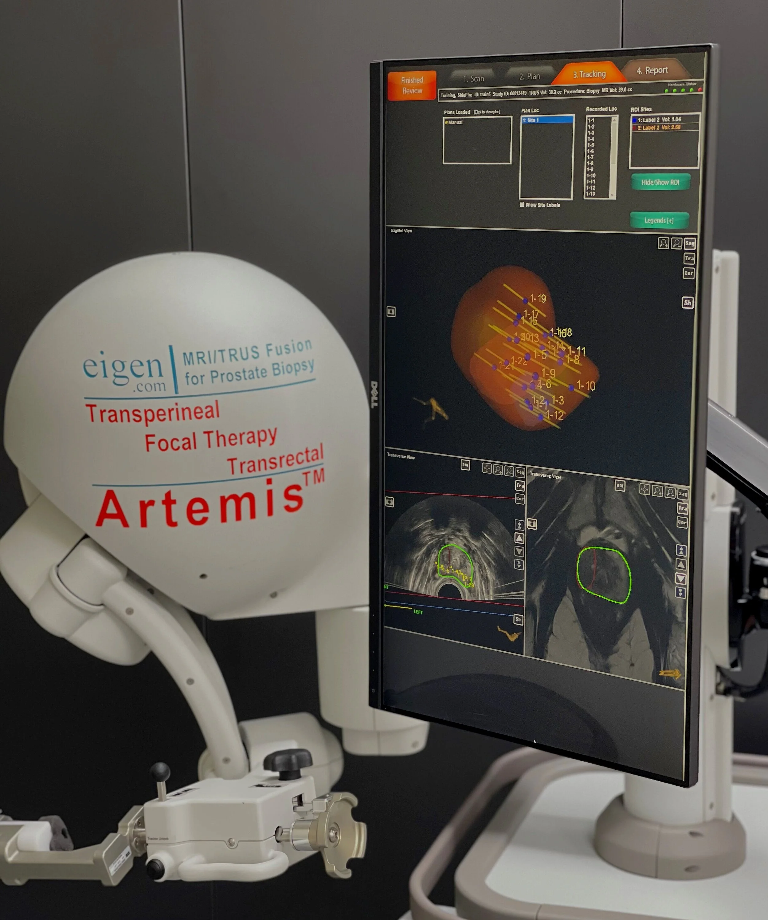

Top: The Red area represents the target region inside the prostate and the lines demonstrate the biopsy samples taken from the prostate
Bottom left: Live Ultrasound of the prostate and computer generated target identification.
Bottom right: Previously obtain MRI with imparted images scan
Artemis System
After an MRI scan of the prostate, abnormal areas or "lesions" may be detected.
Urologists usually use ultrasound to locate the prostate and take biopsies guided by the MRI scan. However, the abnormalities visible on MRI cannot be seen on ultrasound, meaning "cognitive fusion" is used instead. This technique involves estimating the anomaly’s location on the ultrasound and targeting those areas on the prostate. However, this technique is highly dependent on the operator, not reproducible, and prone to error.
Using the ARTEMIS fusion system, we eliminate guesswork from this crucial procedure. We import the MRI images into our specialised ProFuse software, mark the abnormal areas on the MRI, and precisely plan the biopsy locations. These programmed images are then transferred to the ARTEMIS system.
We use ultrasound and specialised software to create a 3D image of the prostate during the procedure. The ARTEMIS system elastically fuses the pre-programmed ProFuse pictures with the live ultrasound 3D reconstructed images, allowing us to identify abnormal areas in real time. The ARTEMIS navigation system then guides our needle to obtain highly accurate and reproducible biopsies of the areas of interest.
All procedures are saved and can be reviewed in the future if necessary.